Сentaur I - Scanning AFM/Confocal/Raman/Fluorescence system for Raman/Fluorescence and AFM/Raman (TERS) imaging
Centaur I - Scanning AFM/Confocal/Raman/Fluorescence system for Raman/Fluorescence and AFM/Raman (TERS) imaging
Centaur combines:
- Scanning Probe Microscope;
- Inverted or Upright Optical Microscope;
- Laser Confocal Microscope;
- Raman Confocal Microscope;
- Fluorescence Confocal Microscope.

Applications:
- Scanning Probe Microscopy;
- Raman Confocal Microscopy;
- Fluorescence Confocal Microscopy;
- Near-Field Scanning Microscopy;
- Tip-Enhanced Raman Spectroscopy (TERS);
- Tip-Enhanced Fluorescent Spectroscopy (TEFS).
Where to use:
- Chemistry. Combination of methods of scanning probe microscopy and Raman spectroscopy allows the analysis of the composition and structure of organic and inorganic substances, traditional and composite materials;
- Physics. Investigation of physical characteristics of surface and subsurface layers of substances and materials;
- Biology. Study of tissues, cells and their structures, biological molecules and the interactions between them;
- Interdisciplinary research. Research in the field of nanotechnology, pharmaceuticals, materials science, mineralogy, geology, forensic, analysis of art and many others.
")
Advantages of Centaur:
- Dual independent scanners (in head and base);
- Multiple simultaneous signal recording (confocal, spectra, topography, phase etc.);
- Full spectra recording in each scan point (hyperspectral imaging);
- Integration with virtually unmodified upright or inverted optical microscopes to work with transparent and none transparent samples;
- Modern cross-platform software (for all the Centaur units).

Components:
- Scanning Probe Microscope Certus;
- XY - sample piezo scanning stage Ratis;
- Confocal unit;
- Monochromator;
- Optical microscope (upright or inverted);
- Digital controller EG-3000;
- Software NSpec.
Raman spectra of carbon nanotubes (CNTs).
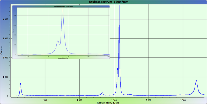
Raman spectra of carbon nanotubes (CNTs).
Grating 1200 lin/mm, exposition time 10 sec, monochromator pinhole 20 μm.
 |
|
 |
 |