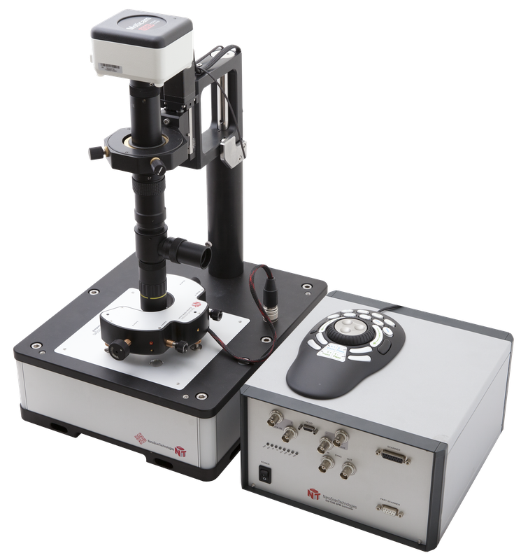
Certus Standard - Basic Configuration of Scanning Probe Microscope
Certus Standard — Basic Configuration of Scanning Probe Microscope
")
Certus Standard - basic configuration of scanning probe microscope, designed to solve a wide range of research and analytical tasks.
Certus Standard includes:
- Scanning head "Certus";
- Video microscope with USB camera;
- Integrated mechanical XY-stage for sample adjustment;
- Digital SPM controller EG-3000;
- NSpec software package;
- Head approach system with 3 motorized actuators.
Certus Standart features:
- Implementation of all basic SPM techniques: Atomic Force Microscopy (AFM, contact and non-contact), shear force AFM, force spectroscopy, Scanning Tunneling Microscopy (STM) etc.;
- Plane-parallel scanning (in X-Y plane) allows imaging with minimal distortion;
- Parallel head approach system;
- Open design of scanning head simplifies observation of the sample and probe at any angle from 0° to 90°;
- Certus Standard is suitable for installation on the optical microscope (upright or inverted), and can also be modified to Certus Optic and Centaur.
")
| A - Certus SPM |
B - Mechanical stage |
| C - Video microscope |
D - CCD camera |
| E - Illumination | F - Vibration protection |
Certus Standard is the best choice for everyday laboratory SPM measurements. Certus Standard could also be interesting to researchers planning to integrate scanning probe microscope with optical and spectral equipment.
|
|
Latex spheres. Image was obtained with SPM Certus Standard. Semi-contact mode. Average sphereas diameter is 94 nm. Image Size 1x1 mkm; 300x300 points. Topography. More detailed information you can find in Latex spheres article. |


SPM head “Certus” contains several probe holders: for standard cantilevers, for “tuning fork” type SPM probes with horizontal and vertical orientation, for STM tips. Any custom design tip holders may be developed by our R&D team by customer request.
One has to change tip holder to change SPM mode.
It is convenient to have several tip holders for use SPM head in cleaned areas, boxes. In this case, only tip holders are being transferred though transition chambers and hatches. You don’t need to move head (SPM microscope).
|
|
The Si/SiO2 periodic structure. Image was obtained with video microscope of SPM Certus Standard. |
|
|
The Si/SiO2 periodic structure and probe. Image was obtained with video microscope of SPM Certus Standard. NSpec window. |
 |
The Si/SiO2 periodic structure and probe. Image was obtained with video microscope of SPM Certus Standart. |
 |
The Si/SiO2 periodic structure. Image was obtained with SPM Certus Standart. Semi-contact mode. Image Size 30x30 mkm; 600x600 points. Topography 3D. More detailed information you can find in Test samples. |


Unique "open design" allows you to use external large aperture objectives, illuminators, microscopes condensers, etc. to illuminate the work area, monitor the sample and the probe position, to take stock of radiation in the point of contact of the probe and the sample.
Certus Standard
Basic datasheet:
|
||||||||||||||||||||||||||||||||||||||||||||||||||||||||||||||||||||||||||||||||||||||||||||||||||||
|
|
||||||||||||||||||||||||||||||||||||||||||||||||||||||||||||||||||||||||||||||||||||||||||||||||||||